The Anatomy of Complex Scoliosis
Understanding Scoliosis begins with understanding anatomy, physics, deformities, and the changes that occur in spinal conditions.
Spinal Anatomy and Complex Scoliosis
Know the parts and pieces that make up the spine. It’s an amazing structure.
A Remarkable Structure
The spine is a stacked column of 24 individual bones called vertebrae.
Vertebrae are separated by fibrous pads of tissue called intervertebral discs. They act as spacers and shock absorbers and allow the spine to bend. Cartilage coats the joint surfaces ensuring smooth movement.
Strong, tough, bands of tissue called ligaments connect the vertebrae, discs and facet joints to help stabilize and support the spinal column at rest and during movement.
When discussing spinal conditions, curve progressions and treatments, your doctor will refer to the spine and vertebrae in sections and numbers. The three sections are Cervical, Thoracic, and Lumbar. An individual or range of vertebrae, like T5-L2. This means the vertebrae between and including the fifth in the Thoracic section through the second in the Lumbar section.
A normal spine looks straight when viewed from the front or back, but has a natural “S” shape when viewed from the side. Scoliosis or Kyphosis are conditions where the spine becomes irregularly curved in one direction or another.
The Scoliotic Curve
The Complex Curve of Scoliosis
The spine curve caused by scoliosis is a result of individual vertebrae tilting and rotating from their original position. The combination of these shifts creates a complex three-dimensional curve.
The curve may occur in the thoracic spine, lumbar spine, or both. Thoracic scoliosis typically curves to the right and lumbar scoliosis typically curves to the left.
Scoliosis is typically diagnosed in adolescents. Most cases are mild, but some curves progress as children grow.
Signs and symptoms of scoliosis may include one shoulder blade being more prominent than the other, uneven shoulders, uneven hips, or one hip being higher than the other.
The Scoliotic Curve
An Unpredictable Path
The curve of the spine that scoliosis causes is often seen as an “S” or “C” shaped curve based on what’s seen on x-ray. Although the exact cause of scoliosis is unknown, the effects are visible and measurable. Changes to the alignment in vertebrae are shifts in both position and rotation, causing the spine to curve in a variety of directions, side to side, forward and back.
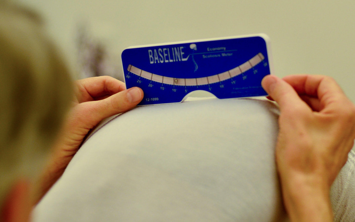
Identifying a curve early is essential to effectively preventing progression. One of the easiest and earliest way to identify a potential curve is called Adam’s Forward Bend Test. It identifies twisting in the spine by seeing the rib cage on one side of the body higher than the other when they are in a bent over position, viewed from behind.
The Cobb Angle
Doctors measure a curve using straight lines and applied geometry.
Angle to Keep an Eye On
How do doctors determine the severity of a scoliotic curve in a standardized way? They define the curve using a measurement called the Cobb angle.
The Cobb angle is a measure of the curvature of your spine in degrees. This measurement helps your doctor determine the progression of a curve by having a standard to compare to over time and to determine what type of treatment is necessary. In order to be diagnosed with complex scoliosis, your Cobb angle must be at least 10 degrees.
The term “progressing” is another way of saying the curve angle is increasing. The Cobb angle is measured using your x-ray. Your medical team determines your Cobb angle by identifying the vertebrae that are most tilted from vertical at both ends of the curve. Lines are drawn across the endplates of those vertebrae. From there, the angle is measured using those lines (see illustration).

